


A magnetic resonance nanoprobe with STING activation character collaborates with platinum-based drug for enhanced tumor immunochemotherapy | Journal of Nanobiotechnology
Supplies and reagents
Manganese chloride (MnCl2), Na2HPO4.12H2O and cyclohexane have been obtained from Aladdin®, China. CO-520 was bought from Sigma-Aldrich, USA. Dioleoyl phosphatidic acid (DOPA), ldl cholesterol, 1,2-dioleoyl-sn-glycero-3-phosphoethanolamine-N-[methoxy(polyethylene, glycol)-2000] and 1,2-dioleoyl-snglycero-3-phosphocholine (DOPC) have been purchased from Avanti Polar Lipids, Inc, USA. Fetal bovine serum (FBS) and Dulbecco’s modified Eagle’s medium (DMEM) have been purchased from Hyclone, USA. The three-(4, 5-dimethyl-thiazol-2-yl)-2,5-diphenyltetrazolium bromide (MTT) was bought from BioSharp (South Korea). All antibodies for movement cytometric evaluation have been purchased from BD Biosciences (USA). cGAS (D3O8O), pSTING(Ser366), STING(D2P2F), pNF-кB p65 (Ser536), NF-кB p65 (L8F6), pIRF3 (Ser396), IRF3 (D83B9), GAPDH have been bought from Cell Signaling Expertise (USA).
Cell strains and animals
MC38 and 4T1 cells have been purchased from Cell Financial institution of Chinese language Academy of Science (Shanghai, China) and have been individually cultured in RPMI and DMEM medium comprising 10% Fetal Bovine Serum (FBS) and antibiotics (1% streptomycin and 100 IU/mL penicillin). Humidified ambiance and 5% CO2 have been wanted for the tradition setting. C57BL/6 (feminine, 5–6 weeks) and BALB/c mice (feminine, 5–6 weeks) have been bought from Hubei Provincial Middle for Illness Management and Prevention (Wuhan, China), and the experimental manipulation on mice was all carried out in line with rules of Animal Care and Use Committee in Huazhong College of Science and Expertise.
Induced polarization of macrophage by Mn2+
Polarization and STING activation of Mn2+ on Uncooked 264.7 cells
Uncooked 264.7 cells have been seeded in a 24-well plate (2 × 10⁵ cells/nicely). After incubation in a single day, the supernatant was discarded. Clean DMEM medium or totally different MnCl2 concentrations (0, 0.1, 0.2, and 0.4 mM) have been added into cells for culturing extra 24 h. Concentrations of IFN-β within the supernatant have been examined by a bioluminescent ELISA package (LumiKine™ Xpress mIFN-β 2.0), and movement cytometer was used to look at the CD86 expression degree of Uncooked 264.7 cells.
To check the ROS manufacturing, Uncooked 264.7 cells have been seeded in a 24-well plate (1 × 10⁵ cells/nicely). After incubation in a single day, the supernatant was discarded. The cells have been washed as soon as with PBS, then clean DMEM or totally different MnCl2 concentrations (0, 0.1, 0.2, and 0.4 mM) have been added into for culturing extra 6 h. The assay package (Beyotime Institute of Biotechnology, China) and movement cytometer was used to check the ROS manufacturing.
STING activation and polarizing bone marrow derived macrophage (BMDM) from M0 to M1 phenotype by Mn2+
BMDM cells have been obtained by referring to earlier strategies [22]. Bone marrow progenitor cells have been extracted from the leg bones of C57 mice, and have been cultured in RPMI-1640 medium that included 20% L929 cultured medium supernatant (vol/vol), 10% FBS, 1% streptomycin and 100 IU mL−1 penicillin. L929 medium may assist to induce bone marrow progenitor cells differentiate into BMDM cells. After culturing for five days, immature BMDM cells (M0) have been collected for additional use.
M0 macrophages have been seeded in a 24-well plate (1 × 10⁶ cells/nicely), and the clean RPMI-1640 or totally different MnCl2 concentrations (0, 50, 100, 200 and 400 μM) have been added for stimulation. After 24 h, the supernatant was collected to check the manufacturing of IFN-β, and the cells have been harvested to look at the CD80 expression degree by a movement cytometer. One other group of BMDM cells (2 × 10⁶ cells/nicely, 6-well plate) that have been handled with MnCl2 (0, 50, 100, 200 and 400 μM) have been lysed to acquire protein, and the expression of cGAS, pSTING/STING, pNF-кB p65/NF-кB p65 and pIRF3/ IRF3 have been examined by western blot.
Polarizing BMDM from M2 to M1 by Mn2+
M0 macrophages have been obtained in line with the procedures described above and seeded in a 24-well plate ((1 × 10⁶ cells/nicely). IL-4 (20 ng/mL) was added and reacted 24 h to induce M0 to M2 phenotype. Subsequent, the medium was changed with totally different concentrations of MnCl2 (0, 50, 100, 200 and 400 μM) to maintain incubating for twenty-four h. Stream cytometer was used to look at the ratio of M1 and M2 macrophages.
Maturation and STING activation of BMDC by Mn2+
BMDC cells have been obtained by referring to earlier strategies [23]. Bone marrow progenitor cells have been extracted from the leg bones of C57 mice, and have been cultured in RPMI-1640 medium that included 20 ng/mL GM-CSF, 10% FBS, 1% streptomycin and 100 IU/mL penicillin. GM-CSF may assist to induce bone marrow progenitor cells differentiate into BMDC cells. After culturing for 7 days, immature BMDC cells have been collected for additional use.
Immature BMDC cells have been seeded in a 24-well plate ((1 × 10⁶ cells/nicely), and LPS and totally different MnCl2 concentrations (0, 50, 100, 200 and 400 μM) in RPMI-1640 medium have been added into for incubation. After 24 h, movement cytometer was used to look at the cell phenotype. Moreover, immature BMDC cells (2 × 10⁶ cells/nicely, 6-well plate) handled with MnCl2 (0, 50, 100, 200 and 400 μM) have been used to check the expression of cGAS, pSTING/STING, pNF-кB p65/NF-кB p65 and pIRF3/ IRF3 by western blot.
STING activation in tumor cells and mouse lung epithelial (MLE) cells by Mn2+
MC38 and MLE-12 cells (5 × 10⁵ cells/nicely, 6-well plate) have been inoculated into 6-well plates. After 12 h, the supernatant was changed by clean medium together with totally different concentrations of MnCl2 (0, 50, 100, 200 and 400 μM) to maintain incubating for twenty-four h. Then cells have been collected and lysed to acquire protein, and the expression of cGAS, pSTING/STING, pNF-кB p65/NF-кB p65 and pIRF3/ IRF3 have been examined by western blot.
MLE-12 cells have been respectively seeded in a 24-well plate (2 × 10⁵ cells/nicely). DMEM medium or totally different MnCl2 concentrations have been added into cells for culturing 24 h. Concentrations of IFN-β within the supernatant have been examined by a bioluminescent ELISA package (LumiKine™ Xpress mIFN-β 2.0).
In vitro cytotoxicity evaluation of Mn2+ in RAW 264.7, BMDC, MC38 and MLE cells
RAW264.7 (2 × 10⁴ cells/nicely), BMDC (2 × 10⁴ cells/nicely), MC38 cells (1 × 10⁴ cells/nicely) and MLE cells (1 × 10⁴ cells/nicely) have been respectively cultured in a 96-well plate for twenty-four h. The supernatant was changed by clean medium together with totally different concentrations of MnCl2 to maintain incubating for twenty-four h. Then the cell viability was examined by MTT assay (λ = 490 nm) utilizing a microplate reader (Multiskan, MK3, Thermo Fisher Scientific, Waltham, MA).
Synthesis and characterization of MnP@Lip
The synthesis process primarily concerned two elements. Half 1, answer A (2 mL CO-520/cyclohexane + 25 μL DOPA (20 mg/mL) + 50 μL Na2HPO4.12H2O (100 mM)) and answer B (2 mL CO-520/cyclohexane + 50 μLMnCl2 (500 mM)) have been stirred individually for 30 min. Below magnetic stirring, answer B was added dropwise into answer A. After stirring for two h, absolute ethanol (4 mL) was used to demulsify. The non-nanoparticles of combined answer have been eliminated by centrifugation (13,000 × g for 15 min). The precipitate (MnP nanoparticles) was collected and dried by nitrogen fuel. Half 2, the phospholipids dissolved in chloroform, consisting of DOPC (40 μL, 20 mM), ldl cholesterol (40 μL, 20 mM) and DSPE-PEG2000 (10 μL, 20 mM), have been added into above precipitate. After the elimination of chloroform by rotary evaporation, phosphate buffer saline (PBS) was added for hydration to kind MnP@Lip nanoparticels.
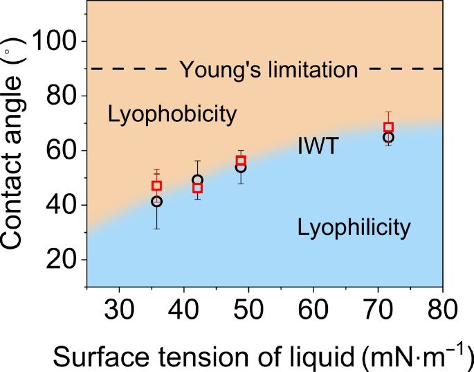
The morphology of MnP nanoparticles and MnP@Lip was confirmed by means of transmission electron microscope (TEM, JEM-1230, Japan). The hydrodynamic diameter, zeta potential and stability of MnP@Lip have been measured and recorded utilizing dynamic gentle scattering (DLS, Zeta Plus, Brookhaven Devices, USA). The discharge conduct of Mn2+ from MnP@Lip in several pH ABS (5.5 and seven.4) have been evaluated utilizing dialysis baggage (molecular weight cutoff, 3500–5000). The pattern exterior the dialysis bag was collected at particular time factors, and the launched quantity of Mn2+ was measured utilizing flame atomic absorption spectroscopy (SpectrAA-240FS, Varian, Palo Alto, CA, USA). The r1 relaxivity of MnP@Lip at totally different pH values (5.5 and seven.4 ABS) was evaluated by a 3 T MRI scanner (123.3 MHz.). The scanning scheme and calculation methodology are according to earlier research [24].
In vivo MR imaging
4T1 orthotopic tumor mannequin was established to confirm in vivo MR imaging skill of MnP@Lip, by utilizing 3 T MRI scanner outfitted with particular mice coil. 4T1 cells (1 × 10⁶/150 μL) have been inoculated to left decrease mammary pad of BALB/c mice, then MnP@Lip (50 μL, 1 mg/kg Mn) was intratumorally injected after 7 days. MR imaging (T1WI) was carried out at pre and 0.5 h, 1 h, 2 h, 5 h, 24 h after MnP@Lip injection. The ratio of tumor and regular tissue sign was calculated to objectively current imaging skill. Detailed MRI scanning parameters are proven in Extra file 1: Desk S1.
Synergistic anti-tumor efficacy research
A number of tumor fashions have been established to validate the synergistic anti-tumor efficacy of MnP@Lip and platinum-based medicine, together with 4T1 orthotopic tumor mannequin, MC38 subcutaneous tumor mannequin and 4T1 incomplete tumor resection mannequin.
4T1 cells (1 × 10⁶/150 μL) have been inoculated to left decrease mammary pad of BALB/c mice to ascertain the orthotopic tumor mannequin. When tumor quantity reached 50–100 mm3, the mice have been randomized into PBS group, MnP@Lip group, oxaliplatin (OXA) group and OXA + MnP@Lip group. Every group of mice acquired matching drug or PBS therapy for 4 instances (with the interval of three days), comparable to PBS (i.t. 100 μL), MnP@Lip (i.t. 1 mg/kg of Mn, 100 μL) and OXA (i.p. 3 mg/kg, 100 μL). Tumor dimension (Quantity = 0.5 × lengh × large2) and weight of the mice throughout the experimentation have been documented each 3 days. On the termination of this analysis, the mice serum was obtained to judge hepatic and renal operate, then tumors have been collected to carry out immunohistochemistry and immunofluorescence assay.
MC38 cells (2.5 × 10⁵/150 μL) have been inoculated to the suitable armpit of C57 mice to ascertain the subcutaneous tumor mannequin. When tumor quantity reached 50–100 mm3, the mice have been randomized into PBS group, cisplatin (Pt) group, and Pt + MnP@Lip group. Every group of mice acquired matching drug or PBS therapy 4 instances (with the interval of three days), comparable to PBS (i.t. 100 μL), Pt (i.p. 3 mg/kg, 100 μL) and MnP@Lip (i.t. 1 mg/kg of Mn, 100 μL). Tumor dimension (Quantity = 0.5 × size × large2) and weight of the mice throughout the experimentation have been documented each 3 days. On the termination of this analysis, tumors have been collected, pictured and weighted.
To ascertain incomplete tumor resection mannequin, 4T1 cells (1 × 10⁶/150 μL) have been inoculated to left decrease mammary pad of Balb/c mice, then tumors have been surgically excised after 10 days of transplantation, and we tried to guarantee that the remaining tumors have been comparable in dimension. The postoperative mice have been randomized into PBS group, OXA group, OXA + MnCl2 group and OXA + MnP@Lip group. Every group of mice acquired matching drug or PBS therapy one time, comparable to PBS (i.t. 100 μL), MnP@Lip (i.t. 1 mg/kg of Mn, 100 μL), MnCl2 (i.t. 1 mg/kg of Mn, 100 μL) and OXA (i.p. 3 mg/kg, 100 μL). The above-mentioned medicine and PBS have been all dispersed in a liquid crystal gel formation system for secure and sustained drug launch in line with earlier research in our lab [25]. The rheology experiments of LCFS precursor and LCFS gel was examined by Dynamic shear rheometer (Kinexus Rotational Rheometer, Malvern Devices, Malvern, UK). In the course of the experimentation, tumor progress and weight of mice have been noticed and recorded. On the termination of this analysis, tumors have been collected to carry out immunohistochemistry and immunofluorescence assay.
In vivo security evaluation
As well as, security research of mixed therapy of MnP@Lip nanoparticles and OXA was carried out. Briefly, PBS, OXA, OXA + MnCl2, and OXA + MnP@Lip have been individually dispersed in LCFS after which they have been individually injected below the pores and skin of the mice. The physique weight of mice was recorded for 7 consecutive days, blood samples have been collected for liver and kidney operate detection, and the HE staining of main tissues and organs have been additionally investigated.
Statistical evaluation
The statistical distinction between teams was evaluated by SPSS software program (model 23.0). The 2-tailed Scholar’s t check or One-way ANOVA (together with Tukey post-hoc check) was used for information satisfying normality and homogeneity of variance. The Mann–Whitney check or Kruskal–Wallis check (together with Bonferroni post-hoc correction) was used to check information that fulfill non-normality or heterogeneity variance. In determine captions, one star (*) means p < 0.05, two (**) means p < 0.01 and three (***) means p < 0.001, and “ns” means no statistically important.